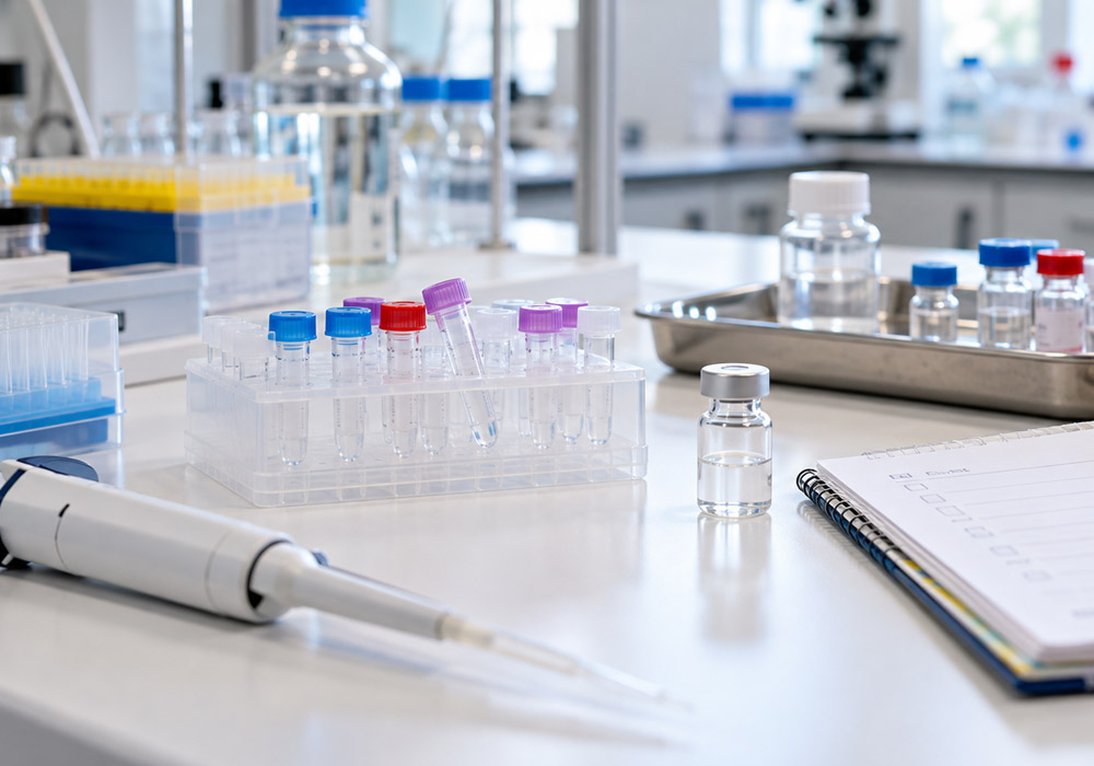
A well-designed peptide experiment can still produce confusing data if material quality goes unchecked or the preparation is incompatible with the assay. Small differences in handling can also make repeat runs difficult to compare. Reviewing the sample from purchase through final concentration calculations helps catch avoidable problems before they obscure the research question.
Assuming the same sequence means equivalent material
Solid-phase peptide synthesis builds chains one residue at a time on resin. Each coupling step can leave behind truncated chains or deletion products. Cleanup removes most of them, but crude material never starts clean. Reversed-phase HPLC is the workhorse for that cleanup, separating the target peak from closely related by-products that would otherwise remain in the vial.
Short peptides can look simple on paper yet involve difficult couplings. Long or hydrophobic stretches can aggregate on resin and produce more synthesis failures. Without a cleanup record, the proportion of target peptide relative to carryover is unclear. That gap can show up later as weak activity or an unexpected dose response. A clean label does not establish clean content.
Two suppliers can list the same sequence and ship different material. Salt form and impurity profile can differ between their preparations. A research-use-only label identifies the intended use, not a universal quality standard. These products are not for human or veterinary use, and their fitness for a laboratory experiment cannot be judged from the sequence line alone.
Sequence provides a starting point, not a guarantee of equivalent material. Batch paperwork and cleanup details help establish what is actually in the vial before an experiment begins.
Skipping a solubility pilot before the full assay
Hydrophobic peptides don’t behave like simple salts in water. They can clump or turn cloudy even when the sequence looks straightforward on paper. That behavior varies with charge and length, so a protocol that worked for one peptide won’t always work for the next. Testing solubility at small scale saves time before committing plates or animals to an experiment.
Start with a small pilot using the solvent named in the supplier paperwork, provided it is compatible with the planned assay. Test dilution into the final assay buffer too: a clear concentrated stock can precipitate after dilution. Watch for haze or particles, but remember that clarity alone does not establish the concentration or exclude small aggregates. If the preparation clouds, pause before scaling up.
DMSO dissolves many sticky sequences, but it can also affect cells when the dose climbs. Dilute acetic acid helps some basic peptides, yet it harms other designs and changes pH. There is no single liquid that suits every peptide, so solvent choice needs to reflect both the sequence and the assay’s tolerance.
Don’t rely on memory alone.
Record what dissolved, at what concentration and at what pH. That preparation note prevents repeat failures when a study scales up or the same stock preparation is repeated.
Filing the certificate of analysis without reading it
The certificate of analysis is a starting point for reproducibility. It should show reversed-phase HPLC data for purity and mass spectrometry data for identity. HPLC area percent expresses the assigned target peak area relative to the total integrated peak area under that method; it does not detect every possible impurity. Mass data supports molecular mass, but intact mass alone doesn’t prove residue order and cannot rule out every isomer. Those limits matter when interpreting the results on the page.
Look past the headline purity number. Review the method details and chromatogram where available rather than relying on the summary alone. Look for peptide content stated separately from HPLC purity, because area purity is not the same as mass of active peptide in the vial. Water and counterions add weight that HPLC percent will not show. Storage advice and appearance notes also matter, since they flag moisture sensitivity or light sensitivity.
Review salt form and terminal details while the sheet is open. A free acid at the C-terminus is not the same molecule as an amidated version. A stated disulfide pattern should line up with the research question and the intended peptide design. If anything is vague, ask for clarification before weighing powder.
A search for “buy peptides Canada” can lead to catalogue options, but availability is only the start of supplier assessment. Use that comparison step to favor lots with full HPLC traces and mass results attached, rather than assessing them by the headline purity figure alone.
Ignoring the counterion and salt form
Trifluoroacetic acid is a common counterion from standard synthesis and cleanup. It stays with the peptide as a salt and can shift pH once the powder dissolves. In sensitive cell systems, that shift can lower viability or alter background signals. The effect depends on the assay, but salt form belongs in the experimental plan.
An alternative counterion, such as acetate, may be worth discussing with the supplier when the existing preparation interferes with an assay. Changing salt form can also change solubility and handling. Neither acetate nor a desalted preparation is automatically suitable for every project. The decision depends on peptide sequence, concentration and the tolerance of the specific readout being used.
Read the certificate to see which salt was received. Check whether the supplier lists residual TFA or acetate content. If the assay is sensitive to pH shifts or counterion load, run a pilot with vehicle controls that contain the same buffer and salt background. Don’t swap salts mid-study without retesting solubility and activity under the relevant assay conditions.
Counterion contributes to the preparation being dosed and should be accounted for accordingly. Recording salt form next to sequence helps later repeats use the same chemistry.
Overlooking storage conditions for powders and solutions
Lyophilization leaves peptides as dry powders that still attract moisture. Once the seal breaks, humidity can start hydrolysis and aggregation even when the vial looks unchanged. Light and warmth add to the damage. Keep unopened vials sealed with desiccant and follow the temperature stated on the certificate rather than choosing storage conditions by habit.
Don’t store dry peptide where temperature swings daily. A freezer door that opens often is a poor spot because cycling brings condensation. A dedicated desiccator or a stable cold spot specified in the paperwork protects the material better. Let a cold vial come to room temperature before opening it, so moist air doesn’t condense on the powder. Weigh quickly and reseal promptly to limit exposure.
Reconstitution creates new risks. A concentrated stock left in the fridge for weeks can lose activity without visible change. Repeated freezing and thawing can increase aggregation or degradation for susceptible sequences, although the effect varies with peptide and formulation. Where frozen storage is allowed by the paperwork, divide fresh solution into single-use aliquots right away.
Thaw one tube per run and discard leftover working solution when the protocol calls for it. That habit costs a few extra tubes, yet it removes a quiet source of drift between otherwise comparable repeats.
Letting oxidation and surface loss go unchecked
Cysteine and methionine need extra care. Free thiols on cysteine can link up and form disulfide artifacts that were never part of the design. Methionine can oxidize, changing shape and binding. Both changes can weaken potency or create mixed populations in the same tube. Visual inspection won’t always reveal the problem, since the solution can remain clear.
Handling deserves attention here. Limit air exposure during weighing and dissolving, and use degassed solvents when the method allows them. When the assay chemistry permits it, a compatible reducing agent such as TCEP can help keep free cysteines reduced. Don’t add it blindly, because some assays react badly to reducers and some peptides are meant to contain defined disulfide bridges.
Intended bridges deserve separate attention. A peptide designed with one or two disulfide bonds is only correct when the pairing matches the target fold. Scrambled pairing gives a different molecule with different activity. Review the stated bridge arrangement and match it to the research question before testing the peptide.
Surface loss is the other silent drain. Hydrophobic sequences can stick to standard plastic tubes or to membranes during sterile filtration. That binding lowers the true concentration in solution while the calculation still assumes full recovery. Low-binding tubes and careful pipetting reduce the problem. Where filtration is needed, assess recovery when possible and keep plastic contact brief throughout preparation.
Calculating from gross weight and overlooking contamination
Gross powder mass can overstate the mass of the target peptide. Counterions, residual water and other non-peptide components contribute weight. Treating all of that weight as the desired molecule can make the actual stock concentration lower than its nominal value and distort a concentration-response comparison.
Use net peptide content when it is stated on the certificate, but read the supplier’s definition. It describes the peptide fraction relative to non-peptide material and can include peptidic impurities; it is not another name for target purity. Both content and purity matter when calculating target concentration. HPLC area purity alone is not an exact mass correction for every mixture. If essential content data are missing, ask the supplier and establish how concentrations will be reported before running quantitative comparisons. Record the lot number, molecular mass and calculation basis so repeats use the same assumptions.
Terminal chemistry needs the same care. An amidated C-terminus and a free acid differ in charge, while a fluorescent tag or biotin handle changes size and binding. None of these versions is wrong on its own, but each answers a slightly different question. Make sure the ordered form matches the form the assay was built around.
Endotoxin deserves attention in sensitive systems. Low-level bacterial fragments can trigger inflammatory signals in cell models and animal research. That background can look like a treatment effect when controls are weak. Where inflammation or immune readouts matter, request assay-appropriate endotoxin data and log those values in the methods. Vehicle controls reveal buffer effects but do not rule out endotoxin carried by the peptide preparation. Use lot testing and additional controls suited to the model to address that possibility.
Peptide work rewards steady habits. Read the paperwork first and test solubility on a small scale before committing material. Respect salt form and the storage limits stated for the lot. Protect sensitive residues and calculate concentrations using true peptide content. Those steps won’t fix a flawed hypothesis, but they help prevent good questions from failing for avoidable handling reasons.